Sacroiliac disease in the horse
Anatomy
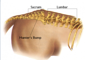
The sacroiliac (SI) joint is a complex region, formed at the junction between the pelvis and the sacrum part of spine. There are several differences between the sacroiliac joint and the other joints in the body; there is only a small amount of joint fluid within the joint space (approximately 1ml); the joint is very stable and very little movement occurs in the region; and the cartilage within the joint differs to other more high motion joints, such as the fetlock. The joint is supported by three sets of ligaments all of which support the SI joint against the weight bearing forces of the horse.
Sacroiliac Disease
Sacroiliac disease refers to any inflammatory process within this region, this can include osteoarthritis, soft tissue injury and occasionally can occur following fractures within the area. As the sacroiliac is difficult to palpate and visualise, even with our diagnostic imaging techniques, conditions are often referred to under the one broad category of sacroiliac disease.
Clinical Signs
The clinical signs suggestive of sacroiliac joint disease can include lameness, a canter quality that is worse than trot, specifically fly kicking out at the canter, croup high canter, breaking from canter back to trot, dragging hind toes and hindlimb stiffness. Often the first subtle clinical sign is non-specific poor performance that you can’t put your finger on.
Diagnosis
The diagnosis of sacroiliac disease is usually made following a thorough clinical examination by your veterinary surgeon ruling out other causes of lameness in the hindlimb(s).
Regional anaesthesia (nerve blocking) of the sacroiliac region can be performed to confirm the diagnosis. This is carried out in a sterile manner by inserting a spinal needle into the region and instilling local anaesthetic. In many cases this is undertaken with the use of ultrasound. The local anaesthetic then diffuses around the region dulling the pain and improving the horses movement, if the horse does indeed have sacroiliac pain.
Quality detailed radiographs (x-rays) of the region are not possible due to the large amount of muscle mass and are rarely, if ever, carried out.
Ultrasonography of the sacroiliac area can be performed and is specifically used to assess the joint capsule and surrounding ligaments. Ultrasonography of this area is not easy and requires some degree of experience to interpret the images accurately. This is done via the skin over the pelvis and also per rectum.
Nuclear Scintigraphy (bone scanning) is currently the most sensitive of the diagnostic imaging techniques. It is not however readily available to most vets and involves a stay at Clyde Vet Group equine hospital, but does incur a higher cost than some of the previously mentioned techniques. It has however been demonstrated that at scintigraphy an increase of the radiopharmaceutical product is only evident in 42% of horses that had a positive nerve block response, demonstrating that there are limitations with this imaging modality. This highlights the importance for diagnosis being multifactorial.
Treatment
Treatment and management of sacroiliac disease is largely nonspecific, this is largely due to the inaccessibility of the joint, which is so well protected by the musculature. The local treatment is achieved in the same manner as the nerve block. Infiltration of the product of choice does not need to be directly into the joint, which is extremely difficult to achieve, but rather relies on the products diffuse around the area therefore as long as the needle is in the correct region. Therapies include peri-articular injections of steroids and occasionally Sarapin®. As with all administration of steroids in the horse, there is a low risk of steroid induced laminitis and we must always be vigilant.
Rehabilitation, in the form of a strict controlled exercise programme alongside physiotherapy is important to help strengthen the surrounding ligaments and musculature to give the sacroiliac joint as much support and strength as possible. The specific programme will be tailored to each horse as an individual with the return to athletic function being at the core of the exercise programme. Systemic non-steroidal anti-inflammatories such as phenylbutazone, also have a part to play in returning the horse to work in as short a period of time as possible.
If you suspect that your horse may have a sacroiliac disease please contact us on 01555 660000.