Injuries to the Deep Digital Flexor Tendon
The deep digital flexor tendon (DDFT) extends from behind the knee and hock, down the back of the cannon, behind the fetlock and pastern joints and ultimately attaches to the underside of the pedal bone within the hoof capsule. The DDFT stabilises the joints of the lower leg when the limb is weight bearing and allows flexion of the digit. The two most common areas for injury of the DDFT are within the hoof capsule or behind the fetlock and pastern which is usually manifest as a windgall (or distension of the digital flexor tendon sheath (DFTS) (figure 1). DDFT damage in the cannon area is very rare.
DDFT injuries within the windgall are more common in hind than forelimbs. They can occur in horses of any age and from a variety of disciplines. Sudden onset, often severe lameness with distension of the DFTS will often be due to a DDFT injury. Ultrasound examination (see fig. 2) will allow the type, extent and severity of the tendon damage to be assessed and a rehabilitation and treatment plan put in place. Treatment will depend on the lesion and can range from rest, along with anti-inflammatories, to medication of the tendon sheath with steroids, to tenoscopy (keyhole surgery) to assess and repair the injury.
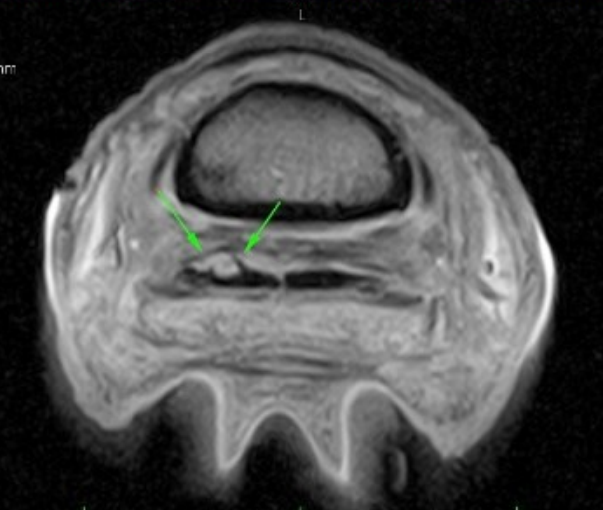
Injuries of the DDFT within the hoof capsule are much more frequent in the forelimb, where they can cause a sudden severe lameness or, more commonly a gradually worsening lameness, often initially only seen on the circle, of one or both limbs. Within the hoof capsule the DDFT runs behind the navicular bone, separated from it by a fluid space, called the navicular bursa. When the horse moves forward the DDFT comes into full contact with the navicular bone and damage in this area can be a major contributor to navicular syndrome. In recent years MRI scans (Fig. 3) have allowed us to further assess lesions to the DDFT within the foot and categorise them. MRIs have allowed a much better ability for vets to advise appropriate management and treatment protocols and give a more accurate prognosis for the likely outcome with each individual horse.
In almost all cases, damage to the DDFT requires a lengthy period of rehabilitation regardless of the treatment approach. This usually involves a period of box rest followed by a slowly ascending exercise program combined with regular reassessments. DDFT injuries are serious and can have significant long-term effects on a horse’s soundness and athletic potential. Careful evaluation of the case, including appropriate imaging techniques, suitable treatment choice, and a well judged rehabilitation plan can enhance the prospect of a return to top performance.
Figure 1: Distension of the digital flexor tendon sheath (windgall). The sudden appearance of this abnormality, coupled with sudden onset lameness, will often indicate damage to the deep digital flexor tendon
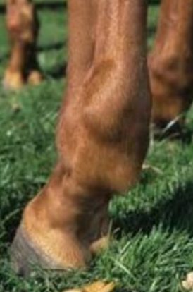
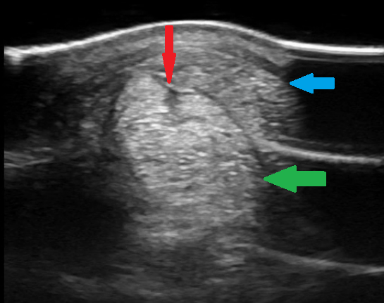
Figure 2. Ultrasound image showing the superficial digital flexor tendon (blue) and the deep digital flexor tendon (green). The red arrow indicates a tear to the deep digital flexor tendon.
Figure 3: MRI showing damage to part of the deep digital flexor tendon with the foot (green arrows). This area should appear similar to the dark area to the immediate right, with sharply defined borders.