Hindlimb Suspensory Injury in Horses.
Ligaments are strong, flexible connective tissue structures that join bone to bone. They are especially important in the biomechanics of the horses’ musculoskeletal system, while also preventing over flexion, extension or rotation of joints. Unfortunately are prone to injury, with the added complication of having poor healing properties, often taking a long time to repair.
The suspensory ligament or ‘interosseous muscle’ is found in both the fore and hindlimbs of the horse. It is made up of bundles of collagen fibres separated by thin layers of a specialised connective tissue. The fibres are arranged in small bundles, which then group to form bigger bundles, much like wire cables used in construction. They are arranged in parallel alignment to each other, running in a longitudinal direction, the same as that of the direction of the force applied to it. This design of the ligament fibres allows it to stretch when loaded, while retaining its strength. The suspensory ligament is an evolutionary derivative of the interosseous medius muscle found in animals with more than one-digit, such dogs or primates. Therefore, muscle fibres are found within the top portion of this ligament, the quantity varying between individual horses and breeds.
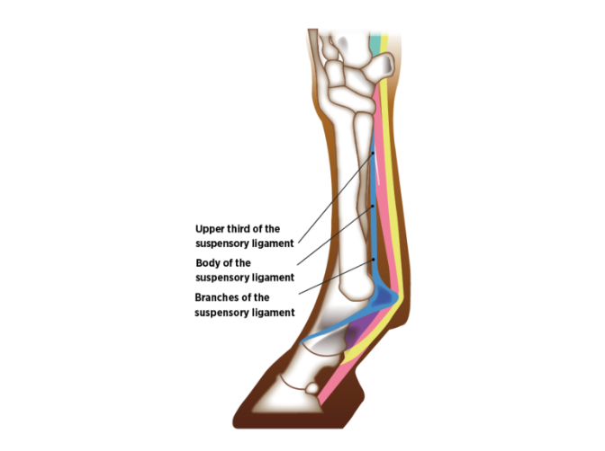
Originating at the top of the back of the cannon bone, between the two splint bones and beneath the flexor tendons, it courses downward towards the fetlock joint. Approximately two thirds of the way down it splits into two branches (the inside (or medial) and the outside (or lateral)) which course over, and attach to, the outside of the respective medial (inside) and lateral (outside) sesamoid bones. It then continues round the front of the pastern to join the main extensor tendon on the front of the limb. The suspensory ligament is also anchored by ligaments that arise at the base of the sesamoids before they insert onto the bones of the pastern.
The main function of the suspensory ligament is to work as part of the ‘suspensory apparatus’ to support the fetlock joint, preventing over extension during loading of the limb while at rest and during exercise. It additionally acts to propel the horse forward during the flight phase of locomotion. It acts like an elastic band, being stretched during the loading phase, building and storing energy, which is then used to propel the horse forward.
It can be seen therefore that a structure that works this hard will be prone to injury and/or wear and tear over time. How does injury happen? Poor hind limb conformation, particularly horses with straight a hock and stifle conformation are prone to hindlimb suspensory problems and this may be associated with weak hyperextending fetlock. Poor hind foot conformation (long toe, low heels) will also increase the risk of damage. Dressage or show jumping are disciplines that increase strain on the suspensory ligament.
Injury can result from either acute overloading or secondary more chronic degenerative changes. Injury occurs when the ligament fibres are stretched excessively, these fibres can then tear, typically at the origin (top of the cannon area) or insertion (branch attachments on to sesamoids). The fibres fray, causing inflammation and bleeding in the affected area. Tearing of the fibres can also occur leaving a focal area of bleeding and swelling, visible on ultrasound and known as a ‘anechoic lesion’. Degenerative changes can occur from repetitive strain injuries; systemic disease such as Pars Pituitary Intermedia Disease (PPID or Cushings disease); or some breed related conditions such as Degenerative suspensory ligament disease (DSLD). DSLD is recognised in old horses and in breeds such as Thoroughbreds, Arabs and Quarter Horses and causes chronic suspensory breakdown and instability with resultant lameness.
Signs of acute or chronic injury to the suspensory ligament may be displayed by lameness, heat or swelling of the affected area. Your vet is likely to diagnose areas of damage with ultrasound imaging. Images can be taken in cross section, across the body and branches of the ligament, and longitudinally, running parallel with the ligament, to assess its structure and presence of an injury. Signs on ultrasound of injury include roughening at the point of origin or insertion, increased cross sectional area and loss of fibre pattern. Ultrasound scanning will also be used to assess the suspensory ligament progression during the treatment and healing process.