A pain in the neck: Neck arthritis in the horse
The equine neck is a complex structure with more than a hundred muscles, tendons and ligaments supporting and moving seven neck bones (cervical vertebrae), extending from poll to withers. The spinal cord spinal cord is protected as it runs through a canal within the middle of these vertebrae, and functions to relay messages from the brain to the peripheral nerves that course throughout the body and back again. Local cervical nerves extend from the spinal cord, through openings at the side of each neck vertebral junction.
Horses, in particular those required to undertake athletic endeavour, are required to use their entire body in their athletic pursuits and the neck plays a vital role in movement, balance and signal transfer to the rest of the body. Unfortunately, successfully diagnosing and treating problem areas can be ‘a pain in the neck’.
Pain or discomfort in the neck results in the horse contracting many of the neck muscles, as an attempt to stabilise the neck, particularly during athletic performance. This can make the entire neck appear stiff and tight and can have longstanding effects including the development of osteoarthritic change.
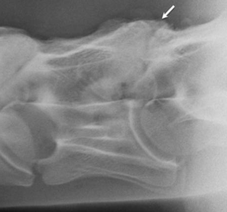
Osteoarthritis or degenerative joint disease is of significant concern for many horses, particularly as they age. Although we often think of this as being a leg and lameness issue, this can also occur in the joints between the cervical vertebrae, the facet joints (Fig. 1). This can result in pain, muscle loss, reduced performance or behavioural issues, as well as forelimb lameness. Many of these signs can be transient and clinically normal individuals can show considerable variation in the appearance of the neck joints on x-ray, making a definitive diagnosis of neck pain challenging. In some 7% or so of horses affected by osteoarthritis of the neck, the spinal cord itself may be affected by excessive new bone formation placing pressure on it. This can result in ataxia (wobbliness / incoordination) which can have repercussions not only for the horse’s ability but also for horse and rider safety.
In order to reach a diagnosis of neck osteoarthritis and commence a treatment plan a number of imaging modalities may be required, in addition to a thorough clinical examination. These may include radiography (x-rays), CT, ultrasound examination and nuclear scintigraphy (‘bone scan’).
Following a diagnosis of neck osteoarthritis several treatment and management options are available. These may include pain relieving anti-inflammatory drugs, steroid medication into the cervical facet joints, guided by ultrasound, a period of time off work or a modified work regime, shock wave therapy and chiropractic adjustments. Whilst the above measures will work, either alone or in combination, in a significant number of horses, the prognosis for neck osteoarthritis is very variable and the ultimate outcome is dependent on many factors including the severity and how advanced the changes are in the joints involved and how much associated soft tissue damage/inflammation there is present.
Despite the difficulties in diagnosing and treating neck pain effectively, new techniques, such as arthroscopic examination of the facet joints and a diagnostic procedure, cervical epiduroscopy, are always evolving, adding to your vet’s ability to manage these cases effectively.
Fig. 1. The white arrow on the radiograph indicates an area of cervical spine osteoarthritis of the facet joint. New bone formation as well as areas of bone resorption and modelling result in the ‘cloudy’, poorly defined margins.

